Our Services

services overview

As a fully licensed and equipped optometric practice, Eyes On J Optometry offers a complete range of eye care services to all our patients.

Whether the eye care issue involves correcting refractive errors with eyeglass or contact lenses, helping a student find amazing frames, or diagnosing/treating eye conditions and diseases, our experienced team will identify and implement the best eye care solutions for you.

Eye Health Evaluation

With our years of experience in diagnosing and treating typical vision disorders such as nearsightedness, farsightedness, amblyopia, presbyopia, cataracts, macular degeneration, and diabetic retinopathy, Sarah Swanson-Harsh O.D., and Spencer Fairgrieve O.D. and their team are equipped to provide appropriate therapeutic medical eye care.

At the same time, the Eyes On J Optometry team offers a wide array of high-quality eye care products at reasonable prices. Our patients never pay too much for the best quality in eyeglasses, contact lenses, sunglasses, progressive and bifocal lenses, and outstanding service.

Treatment of Eye Disease

If you are diagnosed with an eye disease, you want the best treatment available to get your eyes healthy again. At Eyes On J Optometry, we stay current with best treatment practices. Based on your diagnosis, we may recommend a wide variety of approaches, including improved nutrition, prescription medicines, therapy and vision exercises, or medical procedures.

Good Eye Care Begins With A Yearly Eye Exam!

Although many do not realize it, the best way to protect your vision is with a yearly eye checkup.  Even a basic eye exam can instantly detect many health-threatening conditions, such as tumors, vascular irregularities, and diabetes-related injury to the retina. At Eyes On J Optometry, that basic checkup is brief and painless.

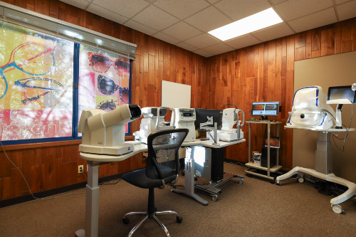
When patients come to our office for eye exams, many times there is testing that we do to help us diagnose problems.  One of these tests uses Optical Coherence Tomography or OCT.   Pictures taken with our OCT machine are generated by light waves that reflect off the back of the eye or retina, creating images similar to what could be produced by a low-power microscope. The OCT also provides cross-sectional images.   These images can display the various layers of the retina.  This technology is also used to image the optic nerve, which is important in glaucoma treatment and management.

OCT PhotoOCT is a non-invasive and no-contact test that doesn't require preparation from the patient. There is no exposure to radiation since the machine uses light to obtain the images.  The patient sits in front of a machine, a couple of bright flashes like a normal camera flash go off, and then the photos can be viewed on the machine within a minute.
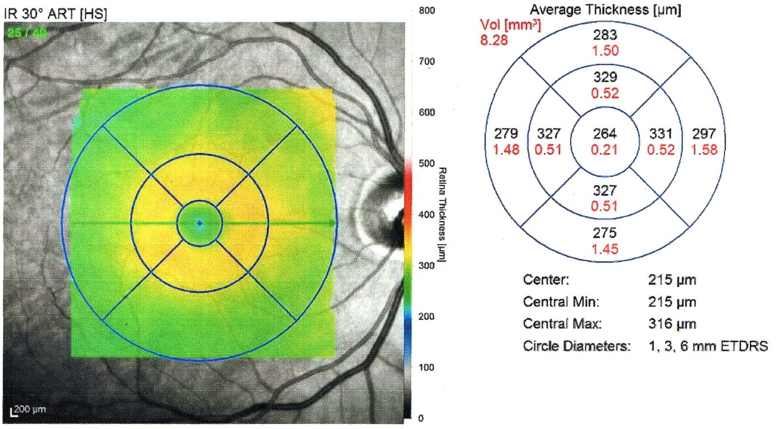
The OCT is an extremely valuable tool used to help diagnose and manage common retinal eye diseases such as macular degeneration, macular edema (fluid in the retina), and macular hole/epiretinal membranes. We use the initial OCT images to aid in making a definitive diagnosis.  The OCT compares these initial images of your eye to a database of images of normal eyes matched to your age. In this way, the first OCT images we take can help point out potential issues if there is something that looks different from normal or average.  Subsequent OCT images can then compare how you look now compared to how you looked initially.  This can be very valuable in gauging how well treatment is working or if the problem is progressing.

OCT Thickness PhotoThe other common use of the OCT is for monitoring and managing glaucoma.  We usually take initial images of the optic nerve, and the OCT can then compare these images to those of age-matched healthy control patients.  The OCT will usually be repeated every year so we can follow any changes or progression over time.
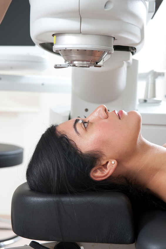
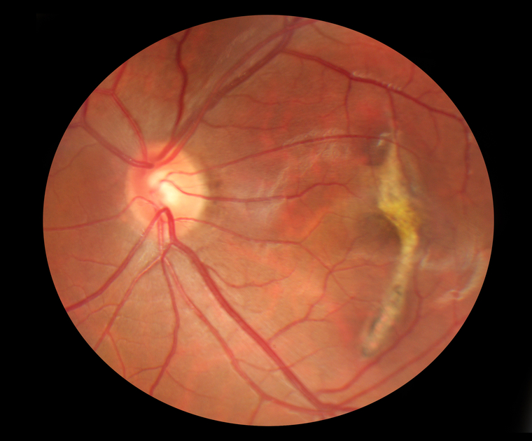
The advent of OCT has revolutionized the way we evaluate the retina because we can now detect subtle findings not otherwise easily seen during clinical exams. This makes the OCT one of the most valuable tests we can do in our office.

lasik
 
LASIK

LASIK, a form of refractive surgery, is an popular option for vision correction, often eliminating the need to wear glasses or contact lenses. Simply put, LASIK reshapes the cornea with a laser.

Other surgical alternatives have become available. Among these is a technique called phakic IOL implantation which involves implanting a lens behind the cornea, but in front of the iris. With this new option, many of those who were too highly nearsighted for LASIK are now candidates for refractive surgery.

If you are interested in refractive surgery, please let us know. Refractive surgery is not to be taken lightly. Detailed testing is necessary to determine whether or not you are a good candidate for the surgery.  If testing shows you to be a good candidate, we can help you choose the refractive surgeon who is most appropriate for your case. In addition, we provide post-operative care for refractive surgery. 

optomap retinal exam
 

In our continued efforts to bring the most advanced technology available to our patients, Sarah Swanson-Harsh O.D., and Spencer Fairgrieve O.D. are proud to announce the inclusion of the Optomap Retinal Exam as an integral part of your eye exam.

Many eye problems can develop without warning and progress with no symptoms. Early on, you might not notice any change in your vision. However, diseases such as macular degeneration, glaucoma, retinal tears or detachments, as well as other health problems such as diabetes and high blood pressure, can often be detected with a thorough exam of the retina. The retina is the part of your eye that catches the image of what you are looking at, similar to the film in a camera.

An Optomap Retinal Exam provides:

  • A scan to confirm a healthy eye or detect the presence of disease.
  • An overview or map of the retina, giving your eye doctor a more detailed view than he can achieve by other means.
  • The opportunity for you to view and discuss the Optomap images of your eye with your doctor at the time of your exam.
  • A permanent record for your medical file, enabling your optometrist to make important comparisons if potential problems show themselves at a future examination.
Interactive Video Player Interactive Video Player

Latest News

Your Eye Floaters and You

Do you have floaters in your vision?

Floaters are caused by thick areas in the gel-like fluid that fills the back cavity of your eye, called the vitreous.

Many people, especially highly near-sighted people, often see some number of floaters for a...

Read more

Brands We Carry

Take a moment to watch the following videos featuring our latest eye health tips, products, and office technology! We welcome you to visit our digital education library as well, which has many more informational videos. If you have questions at any time, be sure to contact us. We'd love to help!


Brands We Carry

Address & Hours

5207 J St Ste 200
Sacramento, CA 95819

(916) 447-3000

Monday 9:00am — 5:00pm
Tuesday 9:00am — 5:00pm
Wednesday 9:00am — 5:00pm
Thursday 9:00am — 5:00pm
Friday 9:00am — 5:00pm
Saturday Closed
Sunday Closed

Contact Us by Email

Directions